

























From Molecular Complementation to Bioluminescence: Principles and Detection Mechanisms of the HiBiT System
In life science research, we often need to answer fundamental questions: How much of a particular protein is expressed? Where is it located within the cell? Which other proteins does it interact with? In situ HiBiT tagging makes protein quantification and functional studies easier. The HiBiT system consists of three main components: The HiBiT tag is derived from NanoLuc luciferase, which originates from the deep-sea shrimp (Oplophorus gracilorostris). After genetic engineering and optimization, NanoLuc luciferase is only 19 kDa and consists of 171 amino acids. It is much smaller than conventional firefly luciferase (approximately 61 kDa), yet substantially brighter—more than 150 times brighter per molecule. Researchers split NanoLuc luciferase into two components. The larger subunit is LgBiT (17.6 kDa), while the smaller peptide is HiBiT, which consists of 11 amino acids and binds strongly to LgBiT. Neither fragment emits light on its own when the substrate furimazine is added. However, when the two fragments come into close proximity, they spontaneously reconstitute the active enzyme and restore its luminescent activity. Figure 1. (a) Comparison of the sizes of NanoLuc and conventional luciferases. (b) Mechanism of HiBiT complementation. Because HiBiT is very small, it has a negligible impact on the target gene and can be readily fused to a protein of interest. Detecting HiBiT therefore enables quantification of the target protein. How can this system be put into practice? Step 1: Insert HiBiT into the target protein at its endogenous locus Step 2: Detect the HiBiT tag Table 1. Two methods for detecting HiBiT-tagged proteins. Step 3: Read the signal Together, these features make HiBiT a sensitive and versatile tool for studying endogenous protein abundance and dynamics. Its small size and high sensitivity make it useful for protein quantification and localization, cell sorting and enrichment, and monitoring protein degradation. However, understanding how to use the system is only part of the equation. A key challenge remains: how can HiBiT be precisely knocked into the endogenous genomic locus using CRISPR? In our next article, we will explore real-world applications of HiBiT and introduce the Flash-KI technology platform, developed specifically to address the challenges of generating knock-in cell lines. In our next article, we will explore real-world applications of HiBiT and introduce the Flash-KI technology platform, developed specifically to address the challenges of generating knock-in cell lines. Learn more about the Flash-KI Platform. Stay tuned! 【1】Hsia, O., et al. Targeted protein degradation via intramolecular bivalent glues. (2024) Nature 627, 204–211.
What Is the HiBiT System?
(1) LgBiT: A 17.6-kDa subunit that has been independently optimized for high structural stability.
(2) HiBiT: An 11-amino-acid peptide with very high affinity for LgBiT (dissociation constant, KD ≈ 700 pM).
(3) NanoLuc substrate (furimazine): Once HiBiT and LgBiT complement one another to form active NanoLuc luciferase, the enzyme catalyzes the oxidation of furimazine. This reaction is ATP-independent. In the presence of oxygen, furimazine is converted to furimamide, releasing carbon dioxide (CO₂) and photons (light).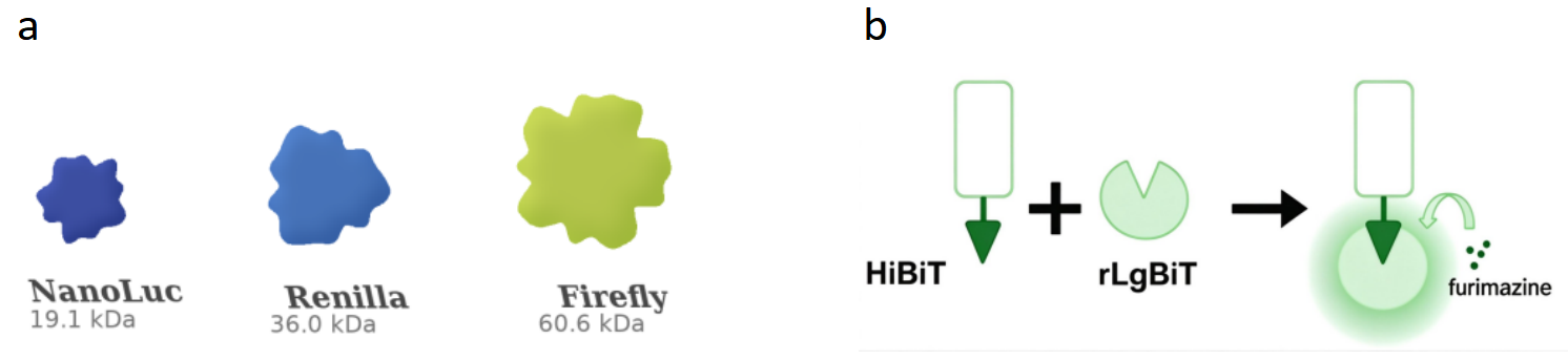
How Is the HiBiT System Used?
To study protein expression and localization under physiological conditions, the CRISPR/Cas9 system can be used to insert HiBiT in situ at the N- or C-terminus of the target protein. The cell then expresses the HiBiT-tagged protein under its native regulatory control. As a result, protein abundance, localization, post-translational modifications, degradation rate, and interactions are maintained as close as possible to their physiological states, making the resulting data more reliable. In certain cases, however, endogenous tagging by genome editing may be difficult. An alternative is to introduce a HiBiT-fusion protein into cells using a plasmid or viral vector. Because this approach relies on an exogenous promoter, it may not reflect physiological expression as faithfully as tagging at the endogenous locus and may produce misleading results.
There are two detection strategies, and the choice depends on the experimental objective:
• LgBiT protein and substrate-based detection: Add a detection reagent containing LgBiT protein and substrate to the engineered cells. Detection can be performed in cell lysates or in live cells. LgBiT binds HiBiT with high affinity, reconstituting the full luciferase enzyme and generating a bioluminescent signal.
• Anti-HiBiT antibody-based detection: Use a monoclonal Anti-HiBiT antibody to recognize the HiBiT tag. This approach uses conventional immunological methods, such as Western blotting, immunofluorescence, flow cytometry, and immunoprecipitation.
Dimension
LgBiT-Based Detection
Antibody-Based Detection
Recognition mechanism
Complementation between HiBiT and LgBiT
Recognition of the tag by an Anti-HiBiT monoclonal antibody
Readout
Bioluminescence
Immunological signals (WB/IF/FACS/IP, etc.)
Typical applications
Protein quantification and screening for protein degradation
Quantification, localization, cell sorting, and enrichment
HiBiT detection is straightforward. Once HiBiT and LgBiT assemble into the active luciferase enzyme, the enzyme emits light on its own and does not require laser excitation. A microplate reader equipped with a luminescence-detection function is therefore sufficient to measure the signal. For protein analysis by Western blot, a chemiluminescence imaging system can be used. To visualize protein distribution in cells, a bioluminescence imaging system can be used.References
【2】Insulin sensitization by small molecules enhancing GLUT4 translocation. Cell Chemical Biology (2023). DOI: 10.1016/j.chembiol.2023.06.012.
【3】Garvin, D. et al. Determining ADCC activity of antibody-based therapeutc molecules using two bioluminescent reporter-based Bioassays. (2021) Curr Protoc. 1(11):e296
Tag
Share


