How Can a Single Base Shape Cortical Folding? CCNB1IP1 Reveals a New Mechanism of Brain Development

Where do the characteristic folds of the cerebral cortex come from? The human cortex has a deeply folded, walnut-like surface, whereas the cortices of many other mammals remain comparatively smooth. Across primate evolution, cortical folding increased gradually rather than appearing all at once through a single "human-specific gene."
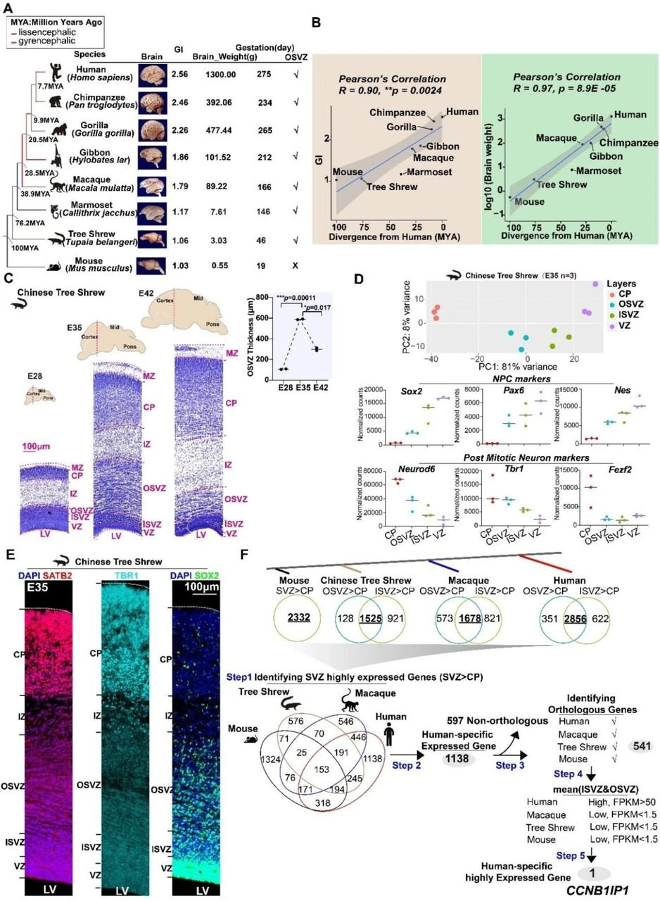
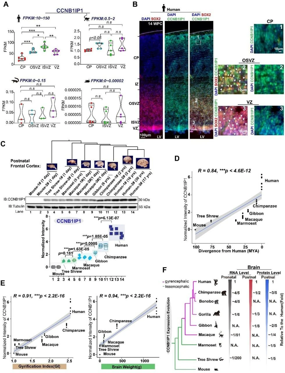
A Nature Genetics study titled "Cis-regulatory evolution of CCNB1IP1 drives the gradual increase of cortical size and folding in primates" focused on CCNB1IP1, a gene that predates primates. Although the gene was already expressed in the brain before primates emerged, its expression rose stepwise across mouse, tree shrew, marmoset, macaque, and human lineages and showed a strong positive correlation with the extent of cortical folding.
CCNB1IP1, short for Cyclin B1 Interacting Protein 1, encodes a RING-domain E3 ubiquitin ligase. Explore the CCNB1IP1 gene page. Its established role in cell biology is to regulate the G2/M phase of the cell cycle by ubiquitinating substrates such as cyclin B.
Traditional explanations for the evolutionary transition from a smooth to a folded cortex have often emphasized new genes that appeared in the human lineage. In this study, however, the researchers found that CCNB1IP1 was already expressed in the tree shrew brain before the emergence of primates, although at a relatively low level.
CCNB1IP1 expression then increased stepwise along the primate lineage. Expression in the smooth-brained marmoset was similar to that in the tree shrew, rose substantially in the folded cortex of the macaque, and reached its highest level in humans. Quantitative analyses showed that CCNB1IP1 expression correlated significantly with brain mass and cortical folding, and closely tracked species divergence time. At the molecular level, the gradual emergence of cortical folds therefore appears to reflect the progressive turning up of an ancient gene's expression rather than the sudden appearance of an entirely new gene.
What drives this stepwise increase in expression? The answer lies not in the protein-coding region, but in nearby cis-regulatory elements. The researchers identified CRE1 as a key regulatory sequence whose activity increased progressively from tree shrews to monkeys, apes, and humans, closely mirroring the rise in CCNB1IP1 expression. Within the core region of CRE1, they pinpointed a decisive single-nucleotide difference: humans carry a G, whereas chimpanzees and other primates carry an A at the corresponding position.
The functional consequence of this single-base difference was tested directly. Changing the human G back to A substantially reduced regulatory activity, whereas converting the chimpanzee A to G increased it. Transcription factor-binding assays suggested that the substitution alters the binding of two transcription factors to CRE1, thereby changing the transcriptional output of CCNB1IP1. Deleting the entire CRE1 element by genome editing also markedly reduced CCNB1IP1 expression, further supporting a causal regulatory role.
This is the central logic behind how a single base can influence cortical folding: the protein itself did not acquire a new function; instead, its regulatory switch was progressively turned up. Evolution does not always depend on the birth of new genes. Small changes in the regulation of existing gene networks can also produce far-reaching morphological effects.
1. Causal Validation
Correlation alone does not establish causation. The most compelling advance in this study was the use of conditional knock-in mice to complete the causal chain.
Using Emx1-Cre and Nestin-Cre, the researchers increased CCNB1IP1 expression in neural progenitor cells to a level comparable to that in macaques. The morphological effect was striking: mice, which normally have a smooth cerebral cortex, developed clear cortical folds, increased brain volume, and a significantly higher local gyrification index. The folding phenotype was reproducible across multiple independent litters. Overall cortical layering was preserved, but upper-layer Satb2-positive and Brn2-positive neurons increased substantially. Their distribution around cortical sulci also resembled patterns observed in gyrencephalic species such as macaques.
At the behavioral level, the knock-in mice showed stronger performance in novel object recognition, increased exploratory activity in the open-field test, and modestly improved learning in the water maze. Raising the expression of this ancient gene was therefore sufficient to initiate a cortical folding program in an otherwise smooth cortex and was accompanied by changes in cognition-related behaviors.
2. Why This Study Matters
- Reframing the origin-of-genes narrative: cortical expansion does not necessarily require human-specific new genes. The cis-regulatory evolution of an ancient, conserved gene can produce major morphological consequences through a gradual increase in expression dosage.
- Explaining evolutionary gradualism: the accumulation of substitutions in regulatory elements can generate stepwise changes in gene expression that parallel the progressive increase in cortical folding across primates.
- Establishing a causal framework: a single noncoding base difference can propagate through cell-cycle regulation, progenitor proliferation, cortical morphology, and learning-related behavior, moving noncoding variation from an association signal toward a testable causal variable.
Cross-species comparisons showed that brain expression of CCNB1IP1 rises stepwise from mouse to tree shrew, marmoset, macaque, and human, and is strongly associated with the extent of cortical folding. The key driver is not a change in the coding sequence, but a single-base substitution in an upstream cis-regulatory element: humans carry G, whereas other primates carry A. G-to-A conversion reduces regulatory activity, while A-to-G conversion increases it. Mechanistically, CCNB1IP1 acts as an E3 ubiquitin ligase that promotes E2F1 degradation, shortens the G1 phase of neural progenitors, stimulates basal progenitor proliferation, increases neuronal output, and supports cortical expansion and folding.
The CCNB1IP1 study illustrates two distinct experimental needs. Demonstrating the regulatory effect of a nucleotide requires precise single-base or multi-base substitution without unintended changes in adjacent sequences. Demonstrating that an expression dosage is sufficient requires controlled knocking in of a species-specific regulatory configuration or coding sequence into a model cell or organism, followed by assessment of the resulting cellular and morphological phenotypes. The first question depends on point-mutation precision; the second depends on targeted integration.
Different cell models can be selected according to the specific question being asked in CCNB1IP1 research:
- Model the CRE1 substitution precisely: EDITGENE's Bingo™ Prime Editing platform can be used to develop custom cell lines carrying the core CRE1 G-to-A or A-to-G substitution, enabling direct comparison of CCNB1IP1 expression and downstream effects across nucleotide states.
- Investigate CCNB1IP1 loss of function: ready-to-use CCNB1IP1 knockout cell lines can support studies of how gene loss affects cell proliferation, cell-cycle progression, and related signaling pathways.
Ready-to-Use CCNB1IP1 Knockout Cell Lines
| Catalog No. | Cell Line | Gene | Gene ID |
|---|---|---|---|
| EDJ-KQ12820 | HEK293 | CCNB1IP1 | 57820 |
| EDJ-KQ41955 | HCT 116 | CCNB1IP1 | 57820 |
| EDJ-KQ40706 | A-549 | CCNB1IP1 | 57820 |
| EDJ-KQ41956 | HeLa | CCNB1IP1 | 57820 |
To model the core CRE1 G-to-A or A-to-G substitution, explore EDITGENE's custom point mutation cell line service; for studies of complete CCNB1IP1 loss of function, select one of the ready-to-use knockout models listed above or request a custom knockout cell line. Contact EDITGENE technical support for model selection, site-feasibility assessment, and a project-specific editing strategy.
References
Follow us on social media





Contact us
+1-833-226-3234 (USA Toll-free)
+1-224-345-1927 (USA)
info@editxor.com
Tag
Share