EDITGENE CO., LTD
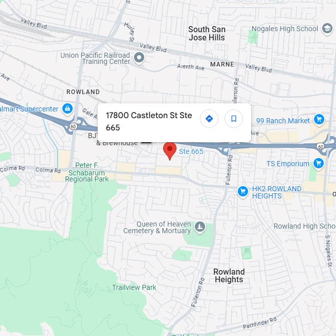
17800 Castleton St. Ste 665. City of Industry. CA 91748
info@editxor.com
+1-833-226-3234 (USA Toll-free)
+1-224-345-1927 (USA)
+86-19120102676 (Intl)

Technical Support

Support Center
Beijing Time: Monday to Friday, 8:00 AM - 6:00 PM
Toll-Free (USA): +833-226-3234
Direct Line (USA): +1-224-345-1927
Email: techsupport@editxor.com

After-Hours Support
Beijing Time: Monday to Sunday, 8:00AM - 6:00 PM
International Line: +86-19120102676
Email: info@editxor.com
Facebook Messenger
Reach out to us on Facebook Messenger for personalized assistance and detailed information.

Linkedin
Engage with us on LinkedIn for professional inquiries, the latest blogs, discoveries, and updates on our innovative work.
FAQ
Which is better for studying MMP25 function, MMP25 Knockout HAP1 Cell Line or MMP25 overexpression HAP1 Cell Line?
The choice depends on whether you are studying MMP25 (MT6-MMP, leukolysin)'s role as a GPI-anchored membrane-type matrix metalloproteinase or its functions in neutrophil biology and emerging cancer roles. The Knockout line is appropriate for asking whether MMP25 is required for these activities — MMP25 is distinct from other MT-MMPs (MMP14/15/16/17/24) in being GPI-anchored rather than transmembrane, and is principally expressed by neutrophils and leukocytes. Overexpression is useful for studying MMP25 in heterologous expression contexts.
Important consideration: MMP25 is principally expressed in neutrophils and leukocytes — HAP1 is not the physiological context for canonical MMP25 functions. The EDITGENE Knockout in HAP1 is most useful for biochemistry, MMP25 substrate identification, and as a clean genetic background for MMP25 structure-function research. Rescue with wild-type or catalytically-dead MMP25 is the standard specificity control. This product complements the parallel MMP15 and MMP16 Knockouts in HAP1 (also available) for MT-MMP family dissection.
What are the application scenarios for this model?
Primary applications:
• Heterologous MMP25 activity: in vitro proteolytic activity assays with candidate MMP25 substrates using recombinant or immunoprecipitated MMP25.
• Surface localization: GPI-anchor topology validation by cell surface staining and PI-PLC sensitivity.
• MT-MMP family comparative studies: parallel analysis with MMP15 and MMP16 Knockouts in HAP1 (also available) for MT-MMP family functional dissection.
• MMP inhibitor specificity: broad-spectrum MMP inhibitor specificity testing in MT-MMP-deficient backgrounds.
EDITGENE recommends this model for in vitro MMP25 biochemistry; physiological neutrophil/leukocyte MMP25 research requires myeloid cell models.
Is this MMP25 Knockout HAP1 Cell Line compatible with overexpression rescue experiments?
Yes. MMP25 rescue experiments require attention to GPI-anchored topology:
• Construct design: use a codon-modified MMP25 sequence with a small N-terminal tag (FLAG, HA) — MMP25 has C-terminal GPI signal that is cleaved during processing, so C-terminal tags will be lost.
• Surface localization validation: confirm GPI-anchored plasma membrane localization by cell surface staining and PI-PLC sensitivity before functional assays.
• Catalytically-dead rescue: zinc-coordinating HEXXH motif glutamate mutation (E to A) abolishes metalloprotease activity and serves as the standard specificity control.
• Functional readout: rescue should restore MMP25-dependent substrate cleavage measured by in vitro proteolytic activity assays.
HAP1-specific considerations:
• Diploidization: HAP1 cells gradually diploidize during extended culture — confirm ploidy by flow cytometry at the time of phenotypic assay.
• Integration site sensitivity: position effects on transgene expression are more pronounced in near-haploid backgrounds; generating multiple independent rescue clones is strongly recommended.
• Transduction efficiency: HAP1 transduces with lentivirus at moderate efficiency — increase MOI compared to standard immortalized lines.
Which is better for studying MAPK15 function, MAPK15 Knockout HAP1 Cell Line or MAPK15 overexpression HAP1 Cell Line?
The choice depends on the experimental question. MAPK15 (ERK7, ERK8) is a less-characterized atypical MAPK with emerging roles in autophagy regulation and ciliary biology. The Knockout line is appropriate for asking whether MAPK15 is required for predicted activities — MAPK15 is unique among MAPKs in having a long C-terminal extension and a TEY activation motif that undergoes autophosphorylation; established functions include phosphorylating LC3-related autophagy proteins and regulating ciliogenesis. Overexpression is useful for studying MAPK15 in heterologous expression contexts.
For atypical MAPK research, the EDITGENE MAPK15 Knockout in HAP1 enables study of this less-characterized member of the MAPK family. Rescue with wild-type or kinase-dead MAPK15 is the standard specificity control. The knockout is valuable for studying MAPK15-dependent autophagy regulation and ciliary biology — MAPK15 has been characterized as a regulator of LC3 trafficking and primary cilium length.
What are the application scenarios for this model?
Primary applications:
• Autophagy regulation: LC3-II accumulation, autophagic flux, and LC3-MAPK15 interaction analysis given MAPK15's role in autophagy.
• Primary cilium biology: ciliogenesis and cilium length analysis given MAPK15's role in cilium regulation.
• ERK7/8 kinase activity: in vitro kinase assays using recombinant or immunoprecipitated MAPK15 with candidate substrates.
• Substrate discovery: phosphoproteomics in the knockout to identify candidate MAPK15-dependent phosphorylation events.
EDITGENE recommends this model for researchers investigating MAPK15 atypical kinase biology, autophagy regulation, and ciliary signaling.
Is this MAPK15 Knockout HAP1 Cell Line compatible with overexpression rescue experiments?
Yes. MAPK15 rescue experiments require attention to unique C-terminal extension:
• Construct design: use a codon-modified MAPK15 sequence with a small C-terminal tag (FLAG, HA). MAPK15 has N-terminal kinase domain with TEY activation motif and unique long C-terminal extension — preserve all elements including the C-terminal regulatory region.
• Kinase-dead rescue: K42R mutation in the ATP-binding lysine abolishes catalytic activity.
• Activation-loop mutant rescue: T175A/Y177F (TEY motif) mutations abolish autophosphorylation-activated kinase activity.
• Functional readout: rescue should restore MAPK15-dependent autophagy regulation and ciliary phenotypes.
HAP1-specific considerations:
• Diploidization: HAP1 cells gradually diploidize during extended culture — confirm ploidy by flow cytometry at the time of phenotypic assay.
• Integration site sensitivity: position effects on transgene expression are more pronounced in near-haploid backgrounds; generating multiple independent rescue clones is strongly recommended.
• Transduction efficiency: HAP1 transduces with lentivirus at moderate efficiency — increase MOI compared to standard immortalized lines.
Which is better for studying MELK function, MELK Knockout HAP1 Cell Line or MELK overexpression HAP1 Cell Line?
The choice depends on whether you are studying MELK (maternal embryonic leucine zipper kinase)'s role as an AMPK-related kinase in cell cycle and stem cell biology or contextualizing it as a historically prominent cancer drug target. The Knockout line is the standard tool for asking whether MELK is required for predicted cellular functions — MELK was initially characterized as a kinase upregulated in many cancers and required for cancer cell proliferation, leading to clinical development of OTSSP167 (a MELK inhibitor). Overexpression is useful for studying MELK gain-of-function effects.
Important historical context: Lin et al. (2017) and Sheltzer et al. demonstrated that MELK CRISPR knockout cancer cells continued to proliferate normally despite MELK loss, contrasting with earlier RNAi-based studies that reported MELK essentiality. OTSSP167 was later shown to have off-target effects accounting for some of its anti-cancer activity. The EDITGENE MELK Knockout in HAP1 provides a genuine genetic null background for rigorous MELK functional studies that complement the older RNAi literature. Rescue with wild-type or kinase-dead MELK is the standard specificity control. The knockout is particularly valuable as a critical specificity control for OTSSP167 and other MELK inhibitors — distinguishing MELK-dependent from off-target effects of these compounds.
What are the application scenarios for this model?
Primary applications:
• OTSSP167 specificity validation: critical genetic control for OTSSP167 (OTS167) — the clinical MELK inhibitor's anti-cancer effects in MELK-null cells reveal off-target contributions distinct from on-target MELK inhibition.
• MELK substrate phosphorylation: phospho-CDC25B and other reported MELK substrates Western blot analysis to characterize MELK kinase activity.
• AMPK-family kinase studies: parallel analysis with AMPK and other AMPK-related kinases for functional specialization characterization.
• Discovery phosphoproteomics: identification of bona fide MELK-dependent phosphorylation events in clean genetic background.
EDITGENE recommends this model for researchers investigating MELK kinase biology and as a critical specificity control for OTSSP167 and emerging MELK inhibitor pharmacology research.
Is this MELK Knockout HAP1 Cell Line compatible with overexpression rescue experiments?
Yes. MELK rescue experiments are well-established for kinase research:
• Construct design: use a codon-modified MELK sequence with a small C-terminal tag (FLAG, HA). MELK has N-terminal kinase domain, UBA (ubiquitin-associated) domain, central regulatory region, and C-terminal KA1 (kinase-associated 1) domain — preserve all elements.
• Kinase-dead rescue: K40R or D150A mutations in the catalytic kinase domain abolish kinase activity and serve as the standard specificity control.
• T-loop phospho-mimetic rescue: T167 (T-loop activation site) E mutation generates constitutively active MELK for gain-of-function studies.
• OTSSP167-resistant rescue: gatekeeper residue mutations (e.g., G89 to large residue) can confer OTSSP167 resistance — useful for confirming on-target inhibitor effects.
• Functional readout: rescue should restore MELK substrate phosphorylation patterns; expectation of restored proliferation depends on context given the CRISPR/RNAi differential dependence finding.
HAP1-specific considerations:
• Diploidization: HAP1 cells gradually diploidize during extended culture — confirm ploidy by flow cytometry at the time of phenotypic assay.
• Integration site sensitivity: position effects on transgene expression are more pronounced in near-haploid backgrounds; generating multiple independent rescue clones is strongly recommended.
• Transduction efficiency: HAP1 transduces with lentivirus at moderate efficiency — increase MOI compared to standard immortalized lines.
Which is better for studying MAP3K9 function, MAP3K9 Knockout HAP1 Cell Line or MAP3K9 overexpression HAP1 Cell Line?
The choice depends on whether you are studying MAP3K9 (MLK1, mixed lineage kinase 1)'s role as a MLK family MAP3K or its functions in JNK pathway activation and stress responses. The Knockout line is the standard tool for asking whether MLK1 is required for these processes — MLK family kinases (MLK1-4, including MAP3K9/MAP3K10/MAP3K11/MAP3K21) activate JNK and p38 pathways downstream of Rac1/Cdc42 GTPases. Overexpression is useful for studying MLK1 gain-of-function effects.
Important consideration: MLK1, MLK2 (MAP3K10), and MLK3 (MAP3K11) share substantial substrate scope — single MLK1 knockout may show modest phenotypes if other MLKs compensate. Rescue with wild-type or kinase-dead MAP3K9 is the standard specificity control. This product complements the parallel MAP3K10 (MLK2) Knockout in HAP1 (also available) for MLK family functional dissection. The knockout is valuable for testing pan-MLK inhibitors (CEP-1347, URMC-099) in neurodegeneration and cancer drug development.
What are the application scenarios for this model?
Primary applications:
• JNK pathway activation: phospho-JNK and phospho-c-Jun following stress stimuli in MAP3K9-null cells.
• Rac1/Cdc42-MLK signaling: assessment of upstream Rac1/Cdc42-driven JNK activation given MLK family GTPase-binding regulation.
• MLK paralog studies: parallel analysis with MAP3K10 (MLK2) Knockout in HAP1 (also available) for MLK family functional dissection.
• MLK inhibitor specificity: critical genetic control for CEP-1347, URMC-099, and other pan-MLK inhibitors in neuroprotection research.
EDITGENE recommends this model for researchers investigating MLK family MAPK upstream activation and MLK-targeted neurodegeneration drug development.
Is this MAP3K9 Knockout HAP1 Cell Line compatible with overexpression rescue experiments?
Yes. MLK1 rescue experiments require attention to MLK family architecture:
• Construct design: use a codon-modified MAP3K9 sequence with a small C-terminal tag (FLAG, HA). MLK1 has N-terminal SH3 domain, kinase domain, leucine zipper (dimerization), CRIB (Rac/Cdc42-binding) motif, and C-terminal regulatory region — preserve all elements.
• Kinase-dead rescue: ATP-binding lysine mutation abolishes catalytic activity.
• CRIB-mutant rescue: CRIB motif mutations disrupt Rac1/Cdc42 binding without affecting intrinsic catalytic activity.
• Functional readout: rescue should restore JNK pathway activation following Rac/Cdc42-activating stimuli.
HAP1-specific considerations:
• Diploidization: HAP1 cells gradually diploidize during extended culture — confirm ploidy by flow cytometry at the time of phenotypic assay.
• Integration site sensitivity: position effects on transgene expression are more pronounced in near-haploid backgrounds; generating multiple independent rescue clones is strongly recommended.
• Transduction efficiency: HAP1 transduces with lentivirus at moderate efficiency — increase MOI compared to standard immortalized lines.
Which is better for studying MSH5 function, MSH5 Knockout HAP1 Cell Line or MSH5 overexpression HAP1 Cell Line?
The choice depends on whether you are studying MSH5's role as a meiosis-specific MutS family member or modeling its associations with premature ovarian insufficiency (POI) and spermatogenic failure. The Knockout line is the standard tool for asking whether MSH5 is required for these processes — MSH5 partners with MSH4 to form MutSγ, a meiosis-specific complex that recognizes Holliday junctions and other meiotic recombination intermediates, distinct from the canonical mitotic MMR functions of MSH2-MSH3/MSH6. Overexpression is useful for studying MSH5 in heterologous expression contexts.
Important consideration: MSH5 is principally functional in meiosis — HAP1 is not the physiological context for canonical MSH5 functions. The EDITGENE MSH5 Knockout in HAP1 is most useful for biochemistry, heterologous expression studies, and as a clean genetic background for MSH5 structure-function research. MSH5 mutations cause autosomal recessive POI 13 (POF13) and have been associated with spermatogenic failure — disease variant rescue enables genotype-function studies. Rescue with wild-type or ATPase-deficient MSH5 enables comprehensive structure-function studies.
What are the application scenarios for this model?
Primary applications:
• Heterologous meiotic studies: in vitro biochemistry of MSH4-MSH5 heterodimer formation and substrate binding.
• POI modeling: rescue with patient-derived MSH5 mutations for genotype-function studies of premature ovarian insufficiency.
• MutSγ assembly: co-immunoprecipitation analysis of MSH4-MSH5 heterodimer integrity.
• Crossover formation studies: in heterologous meiotic-relevant contexts, characterization of MSH5's role in crossover designation.
EDITGENE recommends this model for in vitro MSH5 biochemistry; physiological meiotic MSH5 research requires germline cell models.
Is this MSH5 Knockout HAP1 Cell Line compatible with overexpression rescue experiments?
Yes. MSH5 rescue experiments require attention to MutSγ heterodimer architecture:
• Construct design: use a codon-modified MSH5 sequence with a small C-terminal tag (FLAG, HA). MSH5 has the canonical MutS family architecture with N-terminal mismatch-binding domain, ATPase domain, and dimerization interfaces — preserve all elements.
• ATPase-deficient rescue: ATP-binding lysine mutation abolishes catalytic activity and serves as the standard specificity control.
• MSH4 partnership: MSH5 requires MSH4 for MutSγ formation — rescue interpretation considers MSH4 expression.
• POI mutation rescue: patient-derived MSH5 mutations enable disease genotype-function studies.
• Functional readout: rescue should restore MSH4-MSH5 heterodimer formation; meiotic-specific functions require germline contexts.
HAP1-specific considerations:
• Diploidization: HAP1 cells gradually diploidize during extended culture — confirm ploidy by flow cytometry at the time of phenotypic assay.
• Integration site sensitivity: position effects on transgene expression are more pronounced in near-haploid backgrounds; generating multiple independent rescue clones is strongly recommended.
• Transduction efficiency: HAP1 transduces with lentivirus at moderate efficiency — increase MOI compared to standard immortalized lines.

Room 501, Building D, International Business Incubator, No.3 Juquan Road, Science City, Huangpu District, Guangzhou, Guangdong, China 510663