


-
CRISPR Knockout KitCRISPR Point Mutation KitKI Enhancer Drug
-
Precision Mutation Cell PanelsKnock-in cell lineWild Type Cell Line
-
-
 Cardiovascular - Fluid Shear Stress Response
Fluid shear stress (FSS) is the frictional force generated by blood flow acting on the surface of vascular endothelial cells (ECs) and represents one of the most important mechanical signals in the vascular system. Through sophisticated mechanotransduction mechanisms, endothelial cells convert physical forces into biochemical signals to regulate vascular development and maintain vascular homeostasis. Laminar shear stress promotes anti-inflammatory, anti-thrombotic, and anti-proliferative protective endothelial phenotypes, whereas disturbed flow or low shear stress induces endothelial dysfunction and contributes to the development of cardiovascular diseases such as atherosclerosis.
Fluid shear stress response pathway knockout cell models provide valuable tools for dissecting mechanosensing and signal transduction mechanisms. EDITGENE offers validated knockout cell models targeting key genes in the pathway to support vascular biology research and cardiovascular therapeutic target validation.
The fluid shear stress response pathway describes the key biological processes by which cells or organisms respond to fluid shear stress stimulation. Fluid shear stress is the frictional force generated by blood flow acting on the vascular wall, typically measured in dyn/cm², with physiological arterial shear stress ranging from approximately 10–20 dyn/cm². As the interface between circulating blood and the vessel wall, endothelial cells are continuously exposed to mechanical stimulation and convert physical signals into biochemical signals through specialized mechanosensors, a process known as mechanotransduction.
This pathway regulates endothelial cell alignment, proliferation, migration, and gene expression through activation of multiple mechanosensitive proteins and ion channels, thereby enabling adaptive vascular remodeling during development, tissue repair, and vascular homeostasis. Under physiological conditions, laminar shear stress promotes anti-inflammatory, anti-thrombotic, and anti-proliferative endothelial phenotypes. In contrast, disturbed flow or low shear stress induces endothelial dysfunction and promotes atherosclerotic plaque formation.
However, dysregulated shear stress signaling contributes to multiple diseases. Abnormal shear stress can promote endothelial inflammation, oxidative stress, and apoptosis, driving cardiovascular diseases such as atherosclerosis, restenosis, and hypertension. Due to its critical role in vascular biology and disease progression, the fluid shear stress response pathway has become an important area for cardiovascular disease mechanism research and therapeutic target discovery. Understanding mechanotransduction mechanisms is essential for developing novel therapies targeting endothelial dysfunction.
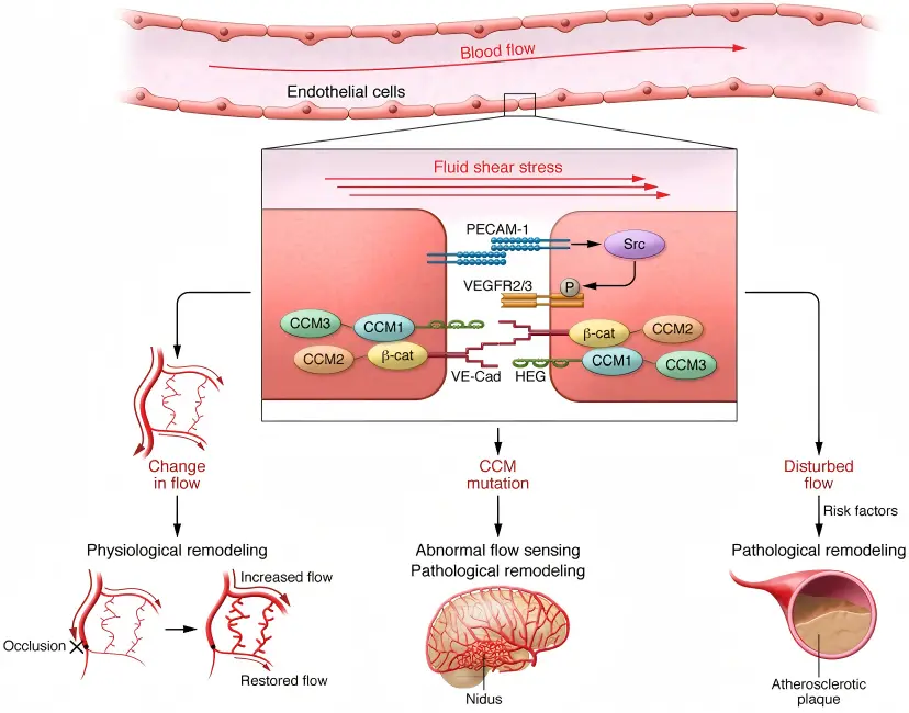
Figure 1. Fluid shear stress sensing in physiological and pathological vascular remodeling.
(Baeyens et al., J Clin Invest, 2016)
Fluid shear stress plays important roles in multiple physiological and pathological processes, including vascular development, atherosclerosis, hypertension, vascular remodeling, and tumor angiogenesis. Dysregulation of key mechanosensors and downstream signaling pathways can enhance endothelial inflammation, oxidative stress, and vascular permeability, contributing to diseases such as atherosclerosis, myocardial infarction, ischemia-reperfusion injury, hypertension, aneurysm formation, and vascular calcification.
These disease contexts further highlight the therapeutic potential of targeting fluid shear stress signaling pathways. Gene knockout cell models provide powerful tools for studying the regulatory functions of key shear stress response genes in different disease settings and support mechanism studies, target validation, and drug discovery.
· Atherosclerosis Models
Used to investigate pro-inflammatory, pro-oxidative stress, and pro-thrombotic endothelial phenotypes under disturbed flow or low shear stress conditions, as well as protective mechanisms induced by laminar shear stress.
· Vascular Development and Remodeling Models
Used to analyze the role of shear stress in embryonic vascular development, arterial-venous differentiation, angiogenesis, and outward vascular remodeling induced by high shear stress.
· Hypertension and Vascular Dysfunction Models
Used to establish endothelial dysfunction models induced by abnormal shear stress, investigate mechanisms underlying vascular stiffness and vasoconstriction/vasodilation imbalance, and evaluate vascular protective agents.
· Tumor Angiogenesis Models
Used to study the effects of abnormal flow patterns within the tumor microenvironment on endothelial cells and investigate the role of shear stress-regulated YAP/TAZ signaling in tumor angiogenesis.
EDITGENE’s fluid shear stress response pathway knockout cell line library includes validated models targeting key regulators of mechanotransduction and vascular homeostasis. We provide high-quality shear stress response knockout cell lines for studying vascular mechanobiology, endothelial dysfunction, and shear stress-related disease pathways. In addition, both ready-to-use and customized knockout cell line services are available to support diverse vascular biology and cardiovascular research needs.
-
Cat.No: EDC90437
Species: Human
Cell Name: HEK293
Gene Name: DRD2
Gene ID: 1813
Specs: 1×10⁶cells
-
Cat.No: EDC90777
Species: Human
Cell Name: A-549
Gene Name: NFE2L2
Gene ID: 4780
Specs: 1×10⁶cells
-
Cat.No: EDC90651
Species: Human
Cell Name: A-549
Gene Name: SMAD7
Gene ID: 4092
Specs: 1×10⁶cells
-
Cat.No: EDC10177
Species: Human
Cell Name: HeLa
Gene Name: GTPBP2
Gene ID: 54676
Specs: 1×10⁶cells
-
Cat.No: EDC07969
Species: Human
Cell Name: HAP1
Gene Name: ABCA1
Gene ID: 19
Specs: 1×10⁶cells
-
Cat.No: EDC07791
Species: Human
Cell Name: HAP1
Gene Name: MAPK7
Gene ID: 5598
Specs: 1×10⁶cells
-
Cat.No: EDC09412
Species: Human
Cell Name: HAP1
Gene Name: TESK1
Gene ID: 7016
Specs: 1×10⁶cells
-
Cat.No: EDC90245
Species: Human
Cell Name: BEAS-2B
Gene Name: PTPN1
Gene ID: 5770
Specs: 1×10⁶cells
-
Cat.No: EDJ-KQ126
Species: Human
Cell Name: HEK293
Gene Name: SMAD6
Gene ID: 4091
Specs: 1×10⁶cells
-
Cat.No: EDJ-KQ223
Species: Human
Cell Name: HEK293
Gene Name: PRKACG
Gene ID: 5568
Specs: 1×10⁶cells
-
Cat.No: EDJ-KQ403
Species: Human
Cell Name: HEK293
Gene Name: SMAD7
Gene ID: 4092
Specs: 1×10⁶cells
-
Cat.No: EDJ-KQ446
Species: Human
Cell Name: HEK293
Gene Name: AKT1
Gene ID: 207
Specs: 1×10⁶cells
-
Cat.No: EDJ-KQ455
Species: Human
Cell Name: HEK293
Gene Name: CSF2
Gene ID: 1437
Specs: 1×10⁶cells
-
Cat.No: EDJ-KQ528
Species: Human
Cell Name: HEK293
Gene Name: SOCS5
Gene ID: 9655
Specs: 1×10⁶cells
-
Cat.No: EDJ-KQ586
Species: Human
Cell Name: HEK293
Gene Name: PTGS2
Gene ID: 5743
Specs: 1×10⁶cells
- 1
- 2
- ...
- 16
- 17
- Next Page »
Subscribe
You can unsubscribe from these communications at any time. For more information on how to unsubscribe, our privacy practices, and how we are committed to protecting and respecting your privacy, please review our Privacy Policy.
By clicking submit below, you consent to allow EDITGENE to store and process the personal information submitted above to provide you the content requested.
