HAP1 Gene-Edited Cell Models: Leveraging Haploid Advantages for Precision Disease Modeling

The HAP1 cell line is derived from the KBM-7 chronic myeloid leukemia (CML) cell line. Through chemical mutagenesis, it has acquired a near-haploid karyotype. While subsequent chromosomal analysis indicates that certain regions may have reverted to diploidy, the overall gene copy number remains simplified, leading to its widespread adoption as a "haploid-like" tool for human genetic screening.
This near-haploid nature implies that most genes exist as a single copy. Once edited via CRISPR, the gene knockout phenotype manifests immediately, thereby bypassing the genetic compensation or interference typically caused by the second allele in diploid cells.
HAP1 has emerged as one of the most popular cell models in functional genetic research. As early as 2015, a research highlight in Nature Reviews Genetics featured genome-wide screening using HAP1. The article, titled "The genetic essence of human cells," spotlighted two independent studies published simultaneously in the journal Science. Both studies utilized haploid cell lines (including HAP1) for genome-wide screening to identify the "core essential genome" of human cells.
01
Why HAP1 is a "Powerful Tool" in Scientific Research
1. Haploid Advantage: Streamlining Gene Editing
The defining characteristic of HAP1 cells is their near-haploid karyotype. With a simplified genomic structure, most genes exist as a single functional copy. Consequently, when utilizing CRISPR/Cas9 for gene knockout, editing efficiency is significantly higher and phenotypic characterization is more direct, drastically shortening experimental timelines.
2. Adherent Growth and Ease of Manipulation
Unlike suspension cells, HAP1 cells exhibit adherent growth and uniform morphology, making them ideal for diverse biochemical assays and microscopic imaging. Their rapid doubling time and ease of expansion make them exceptionally well-suited for large-scale screening experiments.
3. Large-Scale Multi-Omics Studies Validate HAP1’s Potential
In 2022, a landmark study by Rensvold et al., published in Nature, showcased the robust potential of HAP1. The researchers used CRISPR technology to construct 203 mitochondrial gene knockout cell lines in HAP1. Through systematic multi-omics analysis—including proteomics, lipidomics, and metabolomics—they characterized the functions of hundreds of mitochondrial proteins.
This led to the discovery of novel functional genes such as PYURF (NDUFAFQ), SLC30A9, and RAB5IF, while providing a molecular diagnostic basis for patients with undiagnosed mitochondrial diseases. This research underscores the immense value of HAP1 knockout cells in large-scale, systematic functional genomics research.
This led to the discovery of novel functional genes such as PYURF (NDUFAFQ), SLC30A9, and RAB5IF, while providing a molecular diagnostic basis for patients with undiagnosed mitochondrial diseases. This research underscores the immense value of HAP1 knockout cells in large-scale, systematic functional genomics research.
02
Key Application Areas of Gene-Edited HAP1 Cells
The application of gene-edited HAP1 cell lines is exceptionally broad, spanning every level from fundamental cell biology to translational medicine. Below, we illustrate their applications in key research fields using representative gene targets.
1. Kinases and Signal Transduction: From Target Discovery to Drug Resistance Mechanisms
Kinases are the central regulators of cellular signaling networks, and their dysfunction is closely linked to cancer, autoimmune diseases, and metabolic disorders. HAP1 knockout cells provide an ideal platform for dissecting the biological functions of kinases and validating them as therapeutic targets.
● PI3K-AKT Signaling Pathway and Metabolic Reprogramming:
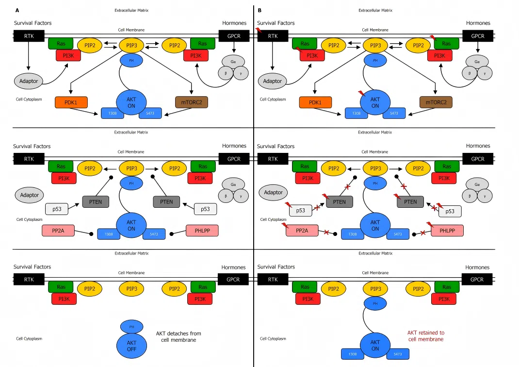
PTEN is a critical negative regulator of the PI3K-AKT signaling pathway, and its loss is a common event in various cancers. The discovery that PTEN deficiency leads to the hyperactivation of mTORC1 is a classic milestone in cancer biology.
By utilizing PTEN knockout HAP1 cells, researchers can clearly recapitulate the hyperactivation of the mTORC1 pathway following PTEN loss. This model allows for in-depth investigations into the regulatory mechanisms of cell proliferation, metabolic reprogramming, and autophagy.
Furthermore, this cell model serves as a vital experimental tool for screening mTOR inhibitors targeting PTEN-deficient tumors and for understanding the metabolic vulnerabilities of cancer cells.
Fig. 1 Biochemical mechanism of PI3K, PTEN and AKT regulation.
● Immunology and the Tumor Microenvironment:
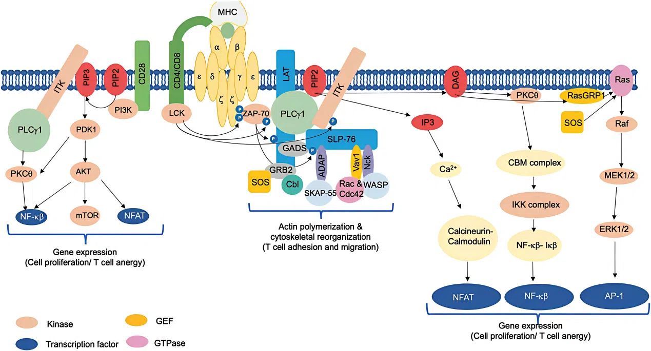
Protein Kinase C theta (PKCθ, encoded by the PRKCQ gene) belongs to the serine/threonine protein kinase family. Its activation is calcium-independent but requires phospholipids as cofactors.
This kinase plays a central regulatory role in T-cell receptor (TCR)-mediated signaling pathways and is a key molecule essential for T-cell activation. Due to its unique position in the immune response, PKCθ has emerged as a significant target in the fields of autoimmune diseases and cancer immunotherapy.
By utilizing PRKCQ knockout HAP1 cells, researchers can precisely evaluate the impact of this kinase's loss-of-function on T-cell signal transduction within an isogenic background. This provides a reliable cell model for drug screening and mechanistic studies targeting PKCθ.
Fig. 2 Positive regulation of T cell signaling.
● Tyrosine Kinases and Neurodevelopment:
The CDKL5 gene encodes a kinase that plays a pivotal role in the development of the nervous system. Pathogenic variants of this gene are the underlying cause of CDKL5 Deficiency Disorder (CDD), a condition clinically characterized by early-onset, refractory epilepsy and severe developmental delays.
Utilizing CDKL5 knockout HAP1 cells allows researchers to investigate the impact of CDKL5 loss on neuronal differentiation, synaptogenesis, and electrophysiological activity within a human cellular context. This model serves as an ideal in vitro platform for dissecting the molecular pathophysiology of CDD and screening for potential therapeutic agents.
2. Epigenetics and Transcriptional Regulation: Unveiling Key Switches in Cancer and Development
Epigenetic mechanisms—including histone modification, DNA methylation, and chromatin remodeling—play a pivotal role in the regulation of gene expression. HAP1 knockout cells provide an ideal experimental platform for investigating the biological functions of these regulatory factors.
● Histone Modification and Gene Transcription:
Lysine Methyltransferase 2C (KMT2C, also known as MLL3) is a histone H3 lysine 4 (H3K4) methyltransferase that catalyzes histone modification marks associated with transcriptional activation. Large-scale cancer sequencing studies have revealed that KMT2C is one of the most frequently mutated epigenetic regulators across various solid tumors, and its loss-of-function (LoF) can promote tumorigenesis and metastasis.
By utilizing KMT2C knockout HAP1 cells, researchers can explore the impact of KMT2C deficiency on chromatin states and gene expression profiles within an isogenic background. This provides a robust cellular model for understanding the tumor-suppressive mechanisms of this gene.
● Super-Enhancers and Oncogene Regulation:
BRD2 belongs to the BET (bromodomain and extra-terminal) protein family. It regulates gene transcription by recognizing acetylated histones and serves as a key factor in super-enhancer regions. BET inhibitors, which function by suppressing BRD2 and other family members to downregulate the expression of super-enhancer-driven oncogenes (such as MYC), currently represent a hot spot in anti-tumor drug development.
By utilizing BRD2 knockout HAP1 cells, researchers can evaluate the impact of BRD2 deficiency on the expression of oncogenes like MYC within an isogenic background. This provides a reliable cellular model for mechanistic studies and efficacy evaluation of BET inhibitors.
3. DNA Damage and Repair: Dissecting Chemotherapy Resistance and Genetic Disease Mechanisms
Defects in DNA damage repair pathways are closely linked to cancer susceptibility, chemotherapy resistance, and various hereditary disorders. Due to their haploid nature, HAP1 cells offer unique advantages in investigating the mechanisms underlying DNA repair.
● The Fanconi Anemia (FA) Pathway and DNA Interstrand Crosslink Repair:
FANCE is a critical component of the Fanconi Anemia core complex. Functional deficiencies in this protein lead to a significant increase in cellular sensitivity to DNA crosslinking agents, such as Mitomycin C (MMC).
In a landmark study, Moder et al. (2017) constructed a FANCC loss-of-function model in HAP1 cells. By combining this with a genome-wide CRISPR library screen, they revealed that the loss of the BLM helicase complex could suppress FANCC-related phenotypes. This study demonstrated that systematic genome-wide screening can effectively identify genetic suppression interactions in the context of DNA repair defects.
The FANCE (c.-56C>T) point mutation HAP1 cell line serves as a powerful tool for investigating the functional nuances of the Fanconi Anemia pathway and for screening synthetic lethal targets.
● Nucleotide Excision Repair (NER) and Chemosensitivity:
ERCC1 is a core factor in the Nucleotide Excision Repair (NER) pathway. It forms a heterodimer with XPF (encoded by the ERCC4 gene) to facilitate the excision of DNA damage induced by UV radiation or chemotherapeutic agents such as cisplatin.
Extensive research has established that the loss of the ERCC1 gene significantly impairs the cellular capacity to repair cisplatin-induced DNA damage, thereby increasing sensitivity to platinum-based drugs, including cisplatin, carboplatin, and oxaliplatin.
Utilizing the ERCC1 (c.*590T>C) point mutation HAP1 cell line and the ERCC4 (c.207+11G>A) point mutation HAP1 cell line, researchers can precisely quantify changes in platinum sensitivity following the loss of these gene functions within an isogenic background. These models are critical for elucidating mechanisms of chemotherapy resistance and developing sensitization strategies.
4. Metabolism and Ion Channels: From Lipid Metabolism to Neuroscience
Metabolic abnormalities and ion channel dysfunctions form the pathological foundation of numerous metabolic and neurological disorders. HAP1 knockout cells provide precision tools for investigating these complex processes.
● Lipid Metabolism and Atherosclerosis:
The Low-Density Lipoprotein Receptor (LDLR) is a critical molecule responsible for clearing LDL cholesterol from the bloodstream. Mutations in the LDLR gene are the primary cause of Familial Hypercholesterolemia (FH). Research has demonstrated that a loss of LDLR function leads to significantly elevated plasma LDL-C levels, which increases the risk of early-onset atherosclerotic cardiovascular disease.
The LDLR (c.*52G>A) point mutation HAP1 cell line enables researchers to study the impact of LDLR deficiency on cholesterol metabolic pathways. This serves as an ideal in vitro cellular model for exploring the molecular mechanisms of Familial Hypercholesterolemia.
● Ion Channels and Neurological Disorders:
Potassium Voltage-Gated Channel Subfamily C Member 1 (KCNC1, encoding the Kv3.1 protein) is highly expressed in fast-spiking inhibitory interneurons and is essential for their high-frequency firing capability.
Whole-exome sequencing (WES) has identified pathogenic missense mutations in the KCNC1 gene that are closely linked to Progressive Myoclonus Epilepsy (EPM7). These mutations cause Kv3.1 channel dysfunction, which impairs the rapid-firing ability of GABAergic interneurons, thereby triggering epileptic seizures.
The KCNC1 knockout HAP1 cell line provides an in vitro research platform to investigate how KCNC1 mutations affect channel function and neuronal electrophysiological properties, facilitating the elucidation of the underlying mechanisms of epilepsy.
5. Ubiquitin-Proteasome System and Apoptosis: Regulating Protein Proteostasis
The Ubiquitin-Proteasome System (UPS) regulates the degradation of the vast majority of intracellular proteins. Its dysfunction is closely linked to neurodegenerative diseases, cancer, and other pathologies. HAP1 knockout cells provide an ideal isogenic model for investigating the functions of UPS-related genes.
● Immunoproteasomes and Antigen Presentation:
PSMB8 encodes the catalytic subunit β5i, a core component of the immunoproteasome. It plays a vital role in intracellular protein degradation, maintenance of homeostasis, and endogenous antigen presentation. Research has revealed that PSMB8 mutations are associated with various autoinflammatory diseases, including Nakajo-Nishimura syndrome and CANDLE syndrome.
By utilizing PSMB8 knockout HAP1 cells, researchers can investigate the impact of immunoproteasome loss-of-function on antigen processing, cytokine production, and inflammatory responses within an isogenic background. This provides a critical cellular model for elucidating the pathogenesis of autoinflammatory diseases and developing targeted therapeutic strategies.
● Deubiquitinating Enzymes and Tumor Suppression:
USP7 (also known as HAUSP) is a critical deubiquitinating enzyme that regulates the stability of p53 and MDM2 proteins through deubiquitination. Research has confirmed that USP7 forms a regulatory complex with p53 and MDM2, playing a central role in maintaining p53 homeostasis.
USP7 is overexpressed in various tumor cells, and inhibiting its activity can effectively suppress tumorigenesis and progression, making it a prominent target in anti-cancer drug development. The USP7 knockout HAP1 cell line provides a reliable in vitro model for screening small-molecule inhibitors targeting USP7 and for investigating the regulatory mechanisms of the USP7-p53/MDM2 axis.
03
HAP1 — EDITGENE Off-the-Shelf Gene-Edited Cell Bank
Core Technologies and Advantages
● Diverse Targets with Comprehensive Coverage
Our off-the-shelf inventory covers a wide range of high-profile research areas, including kinases, epigenetics, DNA damage repair, metabolism, ion channels, neuroscience, autophagy, deubiquitination, and tumor suppression, meeting the diverse needs of the scientific community.
Our off-the-shelf inventory covers a wide range of high-profile research areas, including kinases, epigenetics, DNA damage repair, metabolism, ion channels, neuroscience, autophagy, deubiquitination, and tumor suppression, meeting the diverse needs of the scientific community.
● In-Stock Supply: Ready to Use
Our cell bank maintains a robust inventory, ensuring rapid shipment upon order. This saves you 2-3 months typically required for in-house cell line construction, significantly accelerating your research timeline.
Our cell bank maintains a robust inventory, ensuring rapid shipment upon order. This saves you 2-3 months typically required for in-house cell line construction, significantly accelerating your research timeline.
● Rigorous Quality Control for Reliable Data
Every cell line undergoes comprehensive validation, including Sanger sequencing, mycoplasma testing, and STR profiling. This guarantees accurate cell identity, a contamination-free environment, and highly reproducible experimental results.
Every cell line undergoes comprehensive validation, including Sanger sequencing, mycoplasma testing, and STR profiling. This guarantees accurate cell identity, a contamination-free environment, and highly reproducible experimental results.
● Complete Product Portfolio: A Total Solution
We provide a full suite of products, including Knockout cell lines, Point Mutation cell lines, Overexpression stable lines (Cas9, GFP, Luciferase), and Wild-Type control cells, supporting your entire workflow from gene ablation to functional validation.
We provide a full suite of products, including Knockout cell lines, Point Mutation cell lines, Overexpression stable lines (Cas9, GFP, Luciferase), and Wild-Type control cells, supporting your entire workflow from gene ablation to functional validation.
The complete inventory includes hundreds of gene targets, covering diverse research areas such as kinases, epigenetics, DNA repair, metabolism, and ion channels. For the full product list or to inquire about custom services, please contact EDITGENE Customer Service.
04
Conclusion
From genome-wide CRISPR screens to the functional dissection of disease-related point mutations, and from mitochondrial multi-omics research to the validation of therapeutic targets—HAP1 gene-edited cells, with their unique haploid advantage, have become a cornerstone tool for researchers worldwide to accelerate scientific discovery. The MITOMICS project, published by Rensvold et al. in 2022, has propelled the application of HAP1 cells to new heights of systemic, multi-omic, and high-throughput research.
EDITGENE's off-the-shelf HAP1 cell bank covers hundreds of gene targets. Each cell line is rigorously validated and ready for immediate shipment, helping your research stay ahead of the curve.
References
[1]. Osório, J., et al. (2015). Functional genomics: The genetic essence of human cells. Nature Reviews Genetics, 16(12), 683.
[2]. Rensvold, J. W., et al. (2022). Defining mitochondrial protein functions through deep multiomic profiling. Nature, 606(7913), 382–388.
[3]. Smits, A. H., et al. (2019). Biological plasticity rescues target activity in CRISPR knock outs. Nature Methods, 16(11), 1087–1093.
[4]. Kalaitzidis, D., et al. (2012). mTOR complex 1 plays critical roles in hematopoiesis and Pten-loss-evoked leukemogenesis. Cell Stem Cell, 11(3), 429–439.
[5]. Moder, M., et al. (2017). Parallel genome-wide screens identify synthetic viable interactions between the BLM helicase complex and Fanconi anemia. Nature Communications, 8(1), 1238.
[6]. Leonard, H., et al. (2022). CDKL5 deficiency disorder: clinical features, diagnosis, and management. The Lancet Neurology, 21(6), 563–576.
Contact us
+ 833-226-3234 (USA Toll-free)
+1-224-345-1927 (USA)
info@editxor.com
Tag
Share