Rewriting CRISPR Rules: DNA-Guided Cas12a Enables Precise RNA Cleavage

What if the CRISPR guide is no longer RNA, but DNA? Can the system still work? In the classical CRISPR-Cas12a system, crRNA not only encodes the target sequence information but also stabilizes the active conformation of the Cas protein through its repeat-derived pseudoknot structure.
The PAM sequence on the target DNA serves as the trigger for the activation signal, inducing conformational rearrangement that activates the nuclease function. In this paradigm, the RNA guide is considered an indispensable biochemical prerequisite for Cas protein function.
The research team at the Hong Kong University of Science and Technology posed a question: Can the activation function provided by PAM recognition be physically decoupled from the target information-carrying function of the guide sequence? That is, allowing a DNA molecule to handle the activation function while directing programmable target recognition toward RNA.
The realization of this idea involves designing a synthetic CRISPR DNA (crDNA) that embeds a PAM sequence within its single-stranded sequence and forms a stem-loop structure, allowing it to be recognized by the PI domain of Cas12a and assemble into a functional deoxyribonucleoprotein (DNP) complex. The recognition target of this complex is limited to free RNA molecules.
This discovery was published in Nature Biotechnology under the title “DNA-guided CRISPR–Cas12a effectors for programmable RNA recognition and cleavage.” It completely broke the classical dogma that “CRISPR effectors must rely on RNA guides.”
01
Decoupling Activation and Recognition
The researchers designed a synthetic CRISPR DNA (crDNA) that embeds a PAM and forms a stem-loop structure within its single-stranded sequence. This crDNA can be recognized by the PI domain of Cas12a and assembles into a deoxyribonucleoprotein (DNP) complex.
The DNP complex completely redirects target recognition to free RNA molecules: the activation signal is provided by the DNA guide, while the nucleic acid being recognized and cleaved is RNA.
The cryo-EM structure (3.17 Å) provides direct evidence for this reconstruction.
The crDNA and target RNA form a 20 bp DNA-RNA hybrid duplex that stably binds within the Cas12a substrate channel; the PAM sequence precisely occupies the PI domain in a stem-loop conformation, highly consistent with the PAM-binding mode in classical RNA-guided systems.
The WED-III domain shows increased flexibility in the absence of the RNA pseudoknot, yet the overall architecture of the complex remains similar to that of the natural R-loop. Additional density near the Nuc domain indicates that downstream sequences of the target RNA may loop back into the catalytic cleft for cleavage.
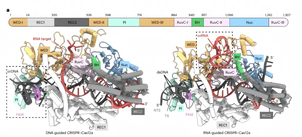 Fig. 1 Structural comparison of DNA-guided and RNA-guided CRISPR–Cas12a systems.
 Figure 1 intuitively demonstrates this reversal: The left side shows the AlphaFold3-predicted model of the DNA-guided ternary complex (crDNA and target RNA forming a hybrid duplex, tightly wrapped by Cas12a’s WED, REC1, and PI domains); the right side shows the classical RNA-guided Cas12a complex with target DNA (PDB 5B43).
The nucleic acid binding modes of the two systems are highly similar, yet the functional logic is completely reversed — the guide changed from RNA to DNA, and the target changed from DNA to RNA.
02
Two-step Activation and Substrate Specificity
Biochemical experiments revealed a non-classical activation pathway: Free Cas12a first binds crDNA through PAM recognition to form the DNP complex (Kd1 = 22.7 nM), then recruits the complementary RNA target to form the ternary complex (Kd2 = 14.2 nM). PAM mutations increased the Kd from 24 nM to 57 nM and reduced cleavage activity, proving that PAM is critical for the first binding step.
The system exhibits strict substrate selectivity: it only recognizes and cleaves ssRNA and shows no detectable interaction with ssDNA or dsDNA. This occurs when the PAM-binding groove is occupied by crDNA, creating steric hindrance that blocks dsDNA access.
The crDNA-ssDNA duplex is unstable within the protein channel and dissociates, as demonstrated by molecular dynamics simulations. Only the DNA-RNA hybrid duplex maintains a stable conformation and triggers activation. Trans-cleavage kinetic shows a kcat approximately half of the RNA-guided system (0.40 vs 0.99 min⁻¹), yet still supporting direct detection of picomolar RNA.
Shortening the crDNA spacer from 20 nt to 16 nt markedly improves single‑base mismatch discrimination — a mismatch at position 12 reduces the signal by ~7‑fold, and at position 15 by ~5‑fold.
03
From Diagnostics to Intracellular RNA Knockdown
Leveraging the stability advantage of DNA guides, the team developed the SLEUTH nucleic acid detection platform, coupling isothermal amplification and T7 transcription with DNA-guided Cas12a trans-cleavage.
It achieves 1 aM detection sensitivity for both RNA and DNA targets and showed complete concordance with RT-qPCR in 31 clinical COVID-19 samples.
In HEK293T cells, unmodified crDNA was ineffective due to rapid degradation. After introducing phosphorothioate (PS) modifications, EGFP fluorescence intensity decreased by ~56% and mRNA levels by 76%, with no effect on non-target transcripts. This represents the first successful specific RNA knockdown using a DNA-guided CRISPR system in mammalian cells.
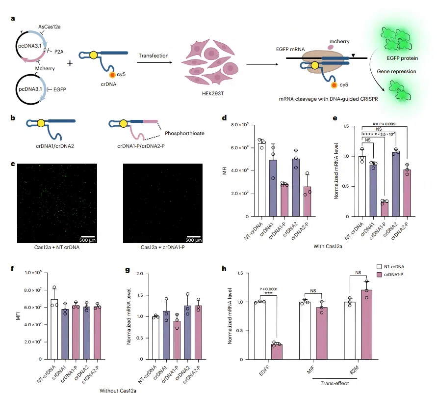 Fig. 2 Intracellular RNA knockdown with DNA-guided CRISPR–Cas system.
 04
Conclusion and Outlook
This Nature Biotechnology study completely breaks the classical understanding that “CRISPR effectors must be guided by RNA” by decoupling the target recognition function of the guide sequence from the allosteric activation function of PAM. The researchers reprogrammed Cas12a into a novel DNA-guided, RNA-targeting effector, marking the first demonstration of programmable RNA knockdown mediated by DNA guides in mammalian cells.
This breakthrough not only highlights the remarkable catalytic plasticity retained by Cas proteins during evolution, but also paves the way for transformative advances in RNA diagnostics, RNA therapeutics, and gene function research.
From attomolar in vitro diagnostics to precise RNA silencing in living cells, the DNA-guided CRISPR-Cas12a system is demonstrating its tremendous potential to advance from basic research to translational applications.
With further optimization of guide structures and effector proteins, this new tool, freed from RNA constraints, is poised to carve out its own unique trail in life sciences and molecular medicine.
Reference
Wu, X., Lam, W.H., Zhao, Z. et al. DNA-guided CRISPR–Cas12a effectors for programmable RNA recognition and cleavage. Nat Biotechnol (2026). DOI: 10.1038/s41587-026-03120-5
Contact us
+ 833-226-3234 (USA Toll-free)
+1-224-345-1927 (USA)
info@editxor.com
Tag
Share